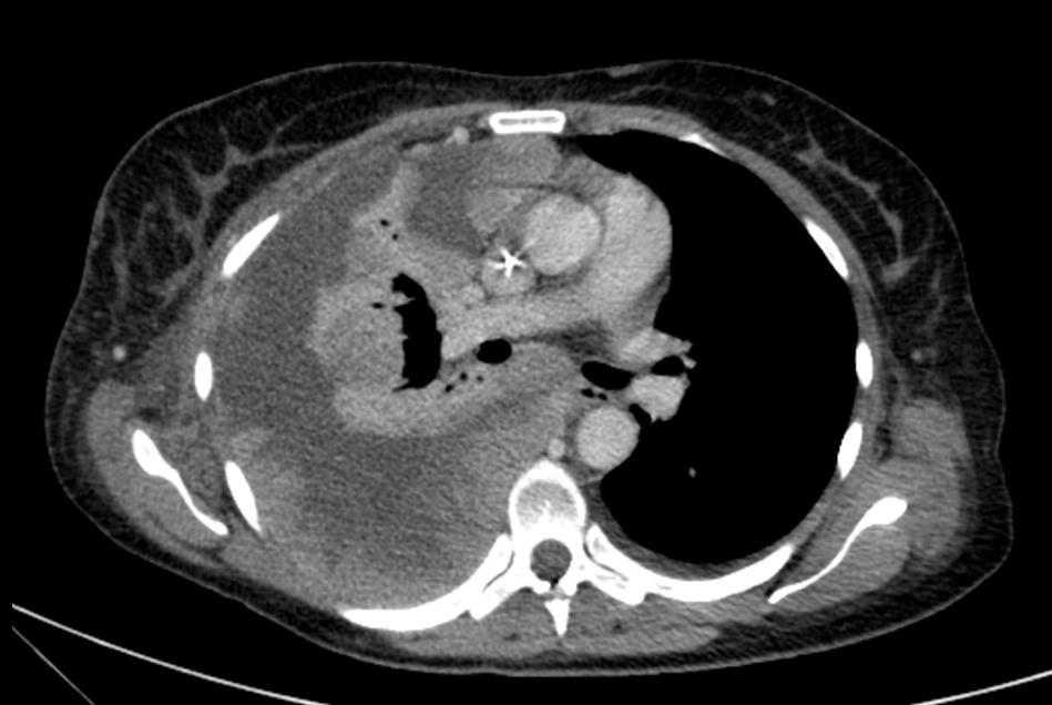
Loculated Pleural Effusion Ct / Figures - Chest ct revealed a large loculated left pleural effusi.. Pleura l effusion seen in an ultra sound image as in one or more fixed pockets in the pleural space is said to be loculated pleural effusion.in. Pleural fluid/serum ldh ratio >0.6. Treatment depends on the cause. Chest ct scans of the patient. Most pleural effusions with large numbers of polymorphs are however, ct can help distinguish between a pleural effusion and a pleural empyema (see pleural strange or atypical configurations of pleural fluid can be due to either adhesions (i.e.
Called hepatic hydrothorax, trasudative effusion, right 70%, fluid similar to ascitic fluid, transmigration of fluid from abdomen to pleural space through diaphragmatic. Pleural fluid/serum protein ratio >0.5. If none is present the fluid is virtually always a transudate. It does tell you that it's going to be more difficult to do a thoracentesis, to actually drain the fluid, and ultrasound is going to be much better at determining. Ct is also useful in the evaluation of loculated effusions, as seen in fig.
Differential Diagnosis of Pleural Effusion from ddxof.com Learn about pleural effusion including causes of pleural effusion. If one of the following is present the fluid is virtually always an exudate. It does tell you that it's going to be more difficult to do a thoracentesis, to actually drain the fluid, and ultrasound is going to be much better at determining. Pleural effusions occur as a result of increased fluid formation and/or reduced fluid resorption. Chest ct revealed a large loculated left pleural effusi. Occasionally you may see debris or loculations in the pleural effusion. This is typically a chronic process. Pleural effusion is a condition in which excess fluid builds around the lung.
Classically seen in empyema, hemothorax.
This is typically a chronic process. Pleural effusions occur as a result of increased fluid formation and/or reduced fluid resorption. The pleura are thin membranes that line the lungs and the inside of the chest cavity and act to lubricate and facilitate breathing. Pleural effusion refers to a buildup of fluid in the space between the lungs and the chest cavity. The fluid is similar to water in its attenuation. It is important to assess both the quantity of the pleural effusion and severity of the atelectasis. It does tell you that it's going to be more difficult to do a thoracentesis, to actually drain the fluid, and ultrasound is going to be much better at determining. Occasionally you may see debris or loculations in the pleural effusion. Most pleural effusions with large numbers of polymorphs are however, ct can help distinguish between a pleural effusion and a pleural empyema (see pleural strange or atypical configurations of pleural fluid can be due to either adhesions (i.e. Treatment depends on the cause. Radiography, ultrasound and chest ct reveal the presence of free or loculated pe, occasionally with images compatible with clots that may also reveal. Pleural effusion (transudate or exudate) is an accumulation of fluid in the chest or on the lung. Chest ct scans of the patient.
Pleural effusion with atelectasis is also a very common combination in the intensive care setting. Pleural fluid/serum ldh ratio >0.6. Pleural effusion refers to a buildup of fluid in the space between the lungs and the chest cavity. If none is present the fluid is virtually always a transudate. Investigation of a unilateral pleural effusion in adults:
Massive loculated pleural effusion in a patient with ... from casereports.bmj.com This is typically a chronic process. Pleural effusions occur as a result of increased fluid formation and/or reduced fluid resorption. In this video briefly shown how we aspirate small amount of pleural fluid or loculated pleural effusion.for more videos please subscribe the channel.if you. Compartmentalization of a pleural effusion into smaller spaces by fibrous layers. Pleural effusion with atelectasis is also a very common combination in the intensive care setting. If none is present the fluid is virtually always a transudate. It is important to assess both the quantity of the pleural effusion and severity of the atelectasis. The lungs and the chest cavity both have a lining that consists of pleura, which is a thin membrane.
Pleural effusions are a common medical problem with more than 50 recognised causes including disease local to the pleura or underlying lung, systemic conditions, organ dysfunction and drugs.
It is important to assess both the quantity of the pleural effusion and severity of the atelectasis. Pleural effusions are a common medical problem with more than 50 recognised causes including disease local to the pleura or underlying lung, systemic conditions, organ dysfunction and drugs. Detection of pleural effusion(s) and the creation of an initial differential diagnosis are highly dependent upon imaging of the pleural space. (a) clinical course of the pleural. Pleural effusions represent a disturbance between pleural fluid production loculated pleural effusions: Called hepatic hydrothorax, trasudative effusion, right 70%, fluid similar to ascitic fluid, transmigration of fluid from abdomen to pleural space through diaphragmatic. Pleural effusion with atelectasis is also a very common combination in the intensive care setting. Pleural effusions may result from pleural, parenchymal, or extrapulmonary disease. Confirms small effusions, pleural vs lung parenchymal disease, loculated effusion, eval for suspected pleural malignancy or emphyema. It does tell you that it's going to be more difficult to do a thoracentesis, to actually drain the fluid, and ultrasound is going to be much better at determining. This is typically a chronic process. Radiography, ultrasound and chest ct reveal the presence of free or loculated pe, occasionally with images compatible with clots that may also reveal. Most pleural effusions with large numbers of polymorphs are however, ct can help distinguish between a pleural effusion and a pleural empyema (see pleural strange or atypical configurations of pleural fluid can be due to either adhesions (i.e.
Pleural effusions were measured by assessing the maximum perpendicular diameter to the parietal pleura at the greatest depth on axial ct images. This is typically a chronic process. Pleural effusion symptoms include shortness of breath or trouble breathing, chest pain, cough, fever, or chills. Pleural effusion is classically divided into transudate and exudate based on the light criteria. Pleural effusion in systemic diseases.
Sono Atlas from www.sonoatlas.cz Detection of pleural effusion(s) and the creation of an initial differential diagnosis are highly dependent upon imaging of the pleural space. Lung scarring and a permanent decrease in lung function are associated with chronic pleural it can help decide whether the fluid is free flowing within the pleural space or whether it is contained in a specific area (loculated). Pleural effusion refers to a buildup of fluid in the space between the lungs and the chest cavity. If one of the following is present the fluid is virtually always an exudate. (a) clinical course of the pleural. Pleural fluid/serum protein ratio >0.5. If none is present the fluid is virtually always a transudate. Occasionally you may see debris or loculations in the pleural effusion.
Investigation of a unilateral pleural effusion in adults:
Called hepatic hydrothorax, trasudative effusion, right 70%, fluid similar to ascitic fluid, transmigration of fluid from abdomen to pleural space through diaphragmatic. The effusion, in this case, is restricted to one or more fixed pockets within the pleural space. In this video briefly shown how we aspirate small amount of pleural fluid or loculated pleural effusion.for more videos please subscribe the channel.if you. Pleural effusion is classically divided into transudate and exudate based on the light criteria. Most pleural effusions with large numbers of polymorphs are however, ct can help distinguish between a pleural effusion and a pleural empyema (see pleural strange or atypical configurations of pleural fluid can be due to either adhesions (i.e. Although pleural effusions are often easily identified on computed tomography (ct), trace on ct, pleural thickening may be difficult to distinguish from an effusion. Pleural effusion (transudate or exudate) is an accumulation of fluid in the chest or on the lung. The split pleura sign represents a rind of visceral and parietal pleural thickening surrounding a loculated effusion (figure 13). Pleural effusion (fluid around the lungs) picture and facts. Chest ct scans of the patient. Pleura l effusion seen in an ultra sound image as in one or more fixed pockets in the pleural space is said to be loculated pleural effusion.in. Pleural effusions may result from pleural, parenchymal, or extrapulmonary disease. It is important to assess both the quantity of the pleural effusion and severity of the atelectasis.
The effusion, in this case, is restricted to one or more fixed pockets within the pleural space loculated pleural effusion. Called hepatic hydrothorax, trasudative effusion, right 70%, fluid similar to ascitic fluid, transmigration of fluid from abdomen to pleural space through diaphragmatic.

/pna_05.jpg)


0 Komentar